
[MD.PhD.] Bordetella pertussis Culture (2026)
Bordetella pertussis Culture: A Laboratory Medicine Specialist’s Complete Guide to Diagnosis and Interpretation

Written by a Board-Certified Laboratory Medicine Specialist (MD.PhD.)
Whooping cough — caused by Bordetella pertussis — is one of the most contagious bacterial respiratory infections known to medicine, and it is making a quiet global comeback. As a Laboratory Medicine Specialist, I frequently see cases delayed in diagnosis because clinicians and patients alike underestimate how deceptively mild the early illness appears, particularly in adults. The B. pertussis culture test remains a cornerstone confirmatory method alongside PCR, offering near-perfect specificity and critical utility for public health surveillance. This post covers everything you need to know: the bacterium’s unique biology, the correct specimen to collect, how the culture is performed, how to interpret results, and where culture fits relative to modern PCR testing.
What Is the Bordetella pertussis Culture Test and Why Is It Ordered?
Bordetella pertussis culture is a microbiological test in which a nasopharyngeal specimen is plated onto specialized growth media and incubated under controlled conditions to isolate and identify the causative agent of pertussis (whooping cough). Because B. pertussis is a fastidious, slow-growing organism with highly specific growth requirements, this is not a routine culture — it demands purpose-built media, careful specimen handling, and extended incubation.
Understanding the pathogen — why it’s so clinically distinct:
B. pertussis is a gram-negative coccobacillus with several defining characteristics that directly impact how the culture test is performed and interpreted:
- Strict aerobe with extremely slow growth kinetics (comparable in fastidiousness to Mycobacterium tuberculosis)
- Exclusively a human pathogen — no animal reservoir
- Produces a distinctive arsenal of toxins that drive the clinical syndrome:
- Pertussis toxin (PT): causes lymphocytosis and systemic immune dysregulation
- Adenylate cyclase toxin: impairs neutrophil and macrophage function
- Tracheal cytotoxin: directly damages ciliated bronchial epithelium, driving the hallmark paroxysmal cough
Clinical indications — when a physician orders this test:
- Confirmation of clinically suspected pertussis in any age group
- Evaluation of prolonged or paroxysmal cough lasting more than two weeks, especially following a prodromal “cold-like” illness
- Outbreak investigation in schools, daycare centers, postpartum care units, or hospitals
- Strain acquisition for epidemiological typing, vaccine efficacy research, and antibiotic resistance surveillance
- Contact tracing and public health reporting in notifiable disease programs
- Evaluation of chronic cough in adults who may be an unrecognized transmission source to unvaccinated infants
The Three Clinical Stages of Pertussis: What Specimen Timing Means for the Lab
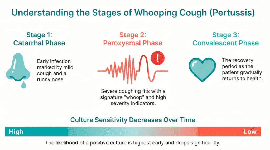
Understanding the disease’s clinical progression is inseparable from understanding when and how to test — because stage of illness at the time of specimen collection is the single most important predictor of culture yield.
| Stage | Duration | Key Features | Culture Yield |
|---|---|---|---|
| Catarrhal (early) | 1–2 weeks | Runny nose, low-grade fever, mild cough | Highest (organism burden at peak) |
| Paroxysmal | 2–6 weeks | Classic “whoop,” post-tussive vomiting; apnea/cyanosis in infants; chronic dry cough in adults | Declining rapidly |
| Convalescent | Weeks to months | Gradually resolving cough | Very low; PCR preferred |
Key insight: Culture sensitivity is at its maximum during the catarrhal stage — often before the diagnosis of pertussis is even considered. By the time the classic whoop is heard, culture sensitivity has already substantially declined. This is why paired PCR testing is essential.
Specimen Collection: The Most Critical Step
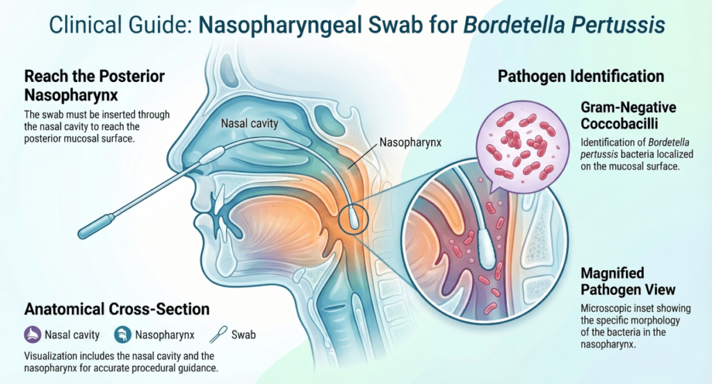
The most consequential variable in B. pertussis culture is not the laboratory — it is the quality and site of specimen collection.
| Specimen Type | Recommendation | Rationale |
|---|---|---|
| Nasopharyngeal (NP) swab | Strongly recommended | Samples the organism’s primary residence in the upper airway mucosa |
| Nasopharyngeal aspirate | Optimal (highest sensitivity) | Larger volume improves organism recovery |
| Sputum | Not recommended | B. pertussis colonizes the nasopharynx, not the lower airway |
| Throat/oropharyngeal swab | Not recommended | Extremely low detection rate |
Collection technique matters: The swab must reach the posterior nasopharynx and be held in place for 10–15 seconds to absorb secretions. Flocked nylon swabs in appropriate transport medium (Regan-Lowe transport medium) are preferred over cotton or alginate swabs, which can be inhibitory to organism viability.
Note: There is no “normal range” for a culture result in the conventional sense — the test is qualitative (positive or negative). The table below summarizes result interpretation rather than a quantitative reference interval.
Culture Result Interpretation
| Result | Interpretation | Recommended Action |
|---|---|---|
| Positive (B. pertussis isolated) | Confirmed pertussis infection | Immediate notification; isolation; macrolide treatment and prophylaxis for contacts |
| Negative (no growth) | Does not exclude pertussis | Correlate with PCR, clinical stage, and specimen quality |
| B. parapertussis isolated | Related but distinct pathogen; causes milder illness | Clinical management similar; notify public health |
| B. holmesii isolated | Rare; associated with immunocompromised hosts | Specialist consultation; confirm with molecular methods |
Note: Reference ranges for quantitative biomarkers vary by laboratory. For culture-based tests, result interpretation must always be made in the full clinical context.
How the Culture Is Performed: Methods and Media
The Culture Method
B. pertussis culture demands specialized conditions that standard bacteriology workflows do not provide.
Recommended growth media:
- Regan-Lowe charcoal agar — current gold standard; contains charcoal (to neutralize fatty acid inhibitors) and 10% horse blood, with cephalexin added to suppress competing nasopharyngeal flora
- Bordet-Gengou agar — the historical medium; still used in some reference laboratories; potato-glycerol-blood base
Incubation conditions:
- Temperature: 35–37°C
- High humidity (desiccation is lethal to this organism)
- Incubation period: minimum 7 days; plates should be held 10–14 days before calling negative
- Examination: colonies appear small (1–2 mm), dome-shaped, glistening — described classically as “mercury droplet” colonies
Identification of isolates:
- Presumptive identification by colony morphology and direct fluorescent antibody (DFA) staining
- Definitive species identification by MALDI-TOF mass spectrometry or PCR-based target gene analysis (IS481 for B. pertussis; IS1001 for B. parapertussis)
Culture vs. PCR: Where Each Method Fits
| Feature | Culture | PCR |
|---|---|---|
| Specificity | ~100% (gold standard for confirmation) | Very high (>95%) |
| Sensitivity | 30–60% (stage-dependent) | 70–99% |
| Optimal timing | Catarrhal stage, pre-antibiotic | Catarrhal and early paroxysmal |
| Turnaround time | 7–14 days | 24–48 hours |
| Post-antibiotic utility | Severely compromised | Remains positive for 3–4 additional weeks |
| Strain available for typing | Yes | No (unless sequenced) |
| Clinical role | Confirmation; outbreak investigation; surveillance | Primary diagnostic test |
Current guideline recommendation (CDC, WHO): PCR is the preferred first-line test for clinical diagnosis. Culture is performed in parallel for public health surveillance, outbreak strain characterization, and epidemiological analysis.
Clinical Significance and Public Health Implications
Confirmed Positive Culture
A culture-confirmed B. pertussis result carries immediate obligations beyond individual patient management:
- Isolation: Infected individuals are contagious from the catarrhal stage until five days after initiation of effective antibiotic therapy
- Treatment: Macrolide antibiotics (azithromycin first-line; clarithromycin or erythromycin as alternatives) shorten the infectious period; they have limited effect on symptom duration if started after the catarrhal stage
- Post-exposure prophylaxis: All household contacts and high-risk contacts (healthcare workers, caregivers of infants) should receive macrolide prophylaxis regardless of vaccination status
- Mandatory reporting: Pertussis is a notifiable disease in most countries; laboratory confirmation triggers formal public health notification and contact investigation
The Under-Recognized Adult as a Transmission Vector
One of the most clinically important — and underappreciated — insights from pertussis epidemiology is that adults with waning vaccine immunity are the primary source of infection for unvaccinated or incompletely vaccinated infants, who bear the highest risk of fatal disease. An adult presenting with a “lingering post-viral cough” of three or more weeks should prompt consideration of pertussis and appropriate testing, even without classic symptoms.
Precautions and Limitations
Factors that reduce culture sensitivity and can cause false-negative results:
- Prior antibiotic use — even a single dose of a macrolide dramatically reduces or eliminates culture yield; PCR sensitivity is also affected but less severely
- Late specimen collection — sensitivity falls sharply after two weeks from symptom onset
- Specimen site error — oropharyngeal or sputum specimens will nearly always yield false-negative results
- Transport delay — B. pertussis is environmentally labile; specimens must reach the laboratory promptly in appropriate transport medium; room-temperature delays of more than two hours reduce viability significantly
- Inhibitory swab materials — cotton or calcium alginate swabs inhibit organism growth
Differentiation from related species:
B. parapertussis and B. holmesii can produce similar clinical syndromes and will not be reliably distinguished by culture morphology alone. MALDI-TOF mass spectrometry or species-specific PCR is required for accurate identification — an important consideration since epidemiological response may differ.
Critical reminder: A negative culture result must never be used as sole justification to exclude pertussis in a clinically compatible case. Culture must always be interpreted alongside PCR results, symptom duration, specimen quality, and antibiotic history. No single laboratory result replaces clinical judgment.
Specialist’s Perspective and Conclusion
In my practice, the most preventable diagnostic failures in pertussis occur at two points: wrong specimen type (oropharyngeal instead of nasopharyngeal) and late collection (paroxysmal stage when the diagnosis is finally considered). If I could embed one message in every clinician’s workflow, it would be this: suspect pertussis early, collect correctly, and always run PCR in parallel with culture.
Culture’s value in 2025 is not primarily speed — PCR wins on that front decisively. Culture’s irreplaceable contribution is providing a viable bacterial isolate that can be characterized genetically and phenotypically. This matters enormously for tracking vaccine-strain divergence, monitoring for macrolide resistance (an emerging concern in some regions), and understanding outbreak dynamics at the population level. Every culture-positive isolate that reaches a reference laboratory contributes to a broader knowledge base that protects future generations.
From a public health standpoint, B. pertussis culture is not just a patient-level diagnostic tool — it is a sentinel surveillance instrument. Clinicians and laboratorians who take it seriously, collect specimens properly, and report results promptly are participating in one of medicine’s oldest and most important public health partnerships.
Key takeaway: Order B. pertussis culture and PCR together, collect from the nasopharynx, do so early in illness, and never let a negative result override strong clinical suspicion.
Author Profile
This article was authored by a board-certified specialist in Laboratory Medicine (MD.PhD.) with expertise in clinical microbiology, infectious disease diagnostics, and public health laboratory science. The author has extensive experience in the laboratory diagnosis of respiratory pathogens and emerging infectious diseases.
[MD.PhD.] Stool Hb Quantitative Test (2026) – MedLab Insight
🧫 Anaerobic Culture Test: A Complete Guide for Clinicians and Patients – MedLab Insight
Some images are generated by AI.
References
UpToDate. (2025). Pertussis infection in adolescents and adults: Clinical manifestations and diagnosis. Wolters Kluwer. https://www.uptodate.com
Centers for Disease Control and Prevention. (2020). Pertussis (whooping cough): Clinical guidance for healthcare professionals. U.S. Department of Health and Human Services. https://www.cdc.gov/pertussis/clinical/index.html
Paddock, C. D., Sanden, G. N., Cherry, J. D., et al. (2008). Pathology and pathogenesis of fatal Bordetella pertussis infection in infants. Clinical Infectious Diseases, 47(3), 328–338. https://doi.org/10.1086/589753
Tatti, K. M., Sparks, K. N., Adkins, B. C., & Tondella, M. L. (2011). Novel multitarget real-time PCR assay for rapid detection of Bordetella species in clinical specimens. Journal of Clinical Microbiology, 49(12), 4059–4066.
World Health Organization. (2018). Pertussis vaccines: WHO position paper — September 2015. Weekly Epidemiological Record. https://www.who.int/docs/default-source/immunization/pertussis_PP_Sep2015.pdf
Loeffelholz, M. J., & Thompson, C. K. (2020). Pertussis diagnosis in the 21st century: Progress and pitfalls. Journal of Clinical Microbiology, 58(7), e00806-19. https://doi.org/10.1128/JCM.00806-19
Murray, P. R., Rosenthal, K. S., & Pfaller, M. A. (2021). Medical microbiology (9th ed.). Elsevier.